Craniometric landmarks
Recently, Caple and Stephan (2015) published an improved classification scheme for anthropometry and a standardized nomenclature for craniometrics points. This scheme is largely based on the sets provided by Martin (1988), with supplements by White (2000), Krogman and Sassouni (1957), Buikstra and Ubelaker (1995) and Howells (1937; 1974), among others. It also takes into account the Bookstein types (I, II or III) (1993) to help elucidate the character and degree of reliability of the points.
Bookstein craniometric landmark types
- Type I landmarks are the most favored, as they tend to be homologous biological points between individuals, are locally defined, and can often be precisely located.
- Type II landmarks are defined by geometric criteria and lack information in at least one direction.
- Type III landmarks are extreme points (often corresponding with instrumentally determined anthropometry), and although the measurements used are reliable and easy to perform, they are often difficult or impossible to establish independently. Furthermore, Type III landmarks depend on the surrounding structures and / or the general orientation of the object, while Type II landmarks are locally defined.
Craniometric landmarks list
| Name | Abbreviation | Type |
|---|---|---|
| Alare | al | III |
| Asterion | ast | I |
| Auriculare | au | II |
| Basion | ba | II |
| Bregma | b | I |
| Coronale | co | III |
| Dacryon | d | I |
| Ectoconchion | ec | II |
| Ectomolare | ecm | III |
| Frontomalare anterior | fma | II |
| Frontomalare orbitale | fmo | II |
| Frontomalare temporale | fmt | III |
| Frontotemporale | ft | II |
| Glabella | g | II |
| Gnathion | gn | III |
| Gonion | go | II |
| Incision | inc | II |
| Infradentale | id | II |
| Inion | i | II |
| Lamda | l | I |
| Mastoidale | ms | II |
| Maxillofrontale | mf | I |
| Menton | me | III |
| Mid-supraorbital | mso | II |
| Nasion | n | I |
| Ophistion | o | II |
| Opisthocranion | op | III |
| Orbitale | or | II |
| Pogonion | pg | III |
| Porion | po | II |
| Prosthion | pr | II |
| Rhinion | rhi | I |
| Subspinale | ss | II |
| Supramentale | sm | II |
| Vertex | v | III |
| Zygion | zy | III |
| Zygomaxillare | zm | III |
| Zygoorbitale | zo | II |
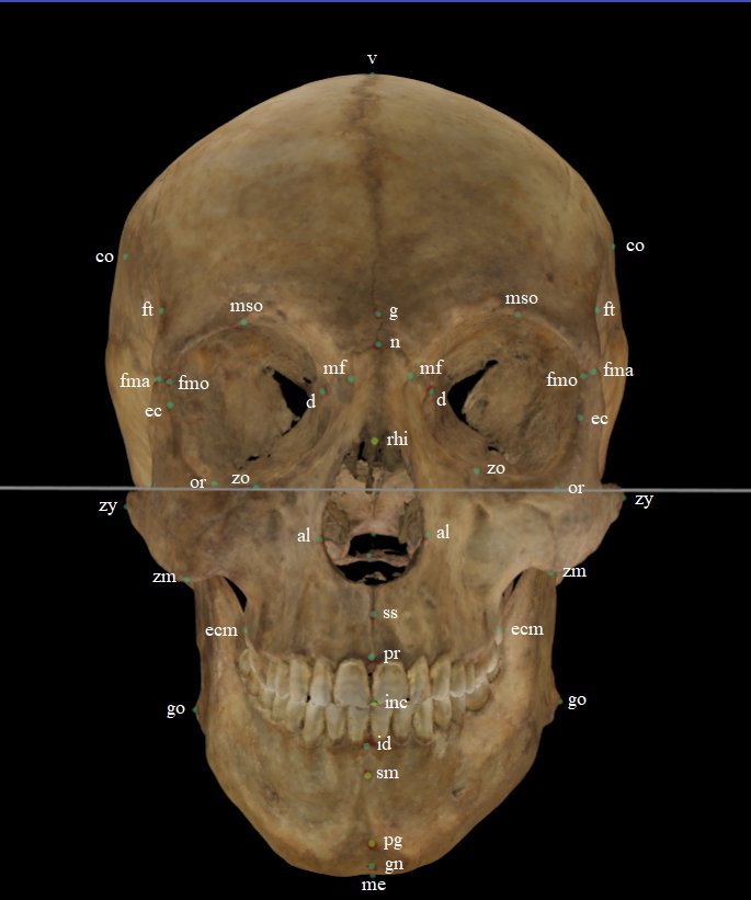
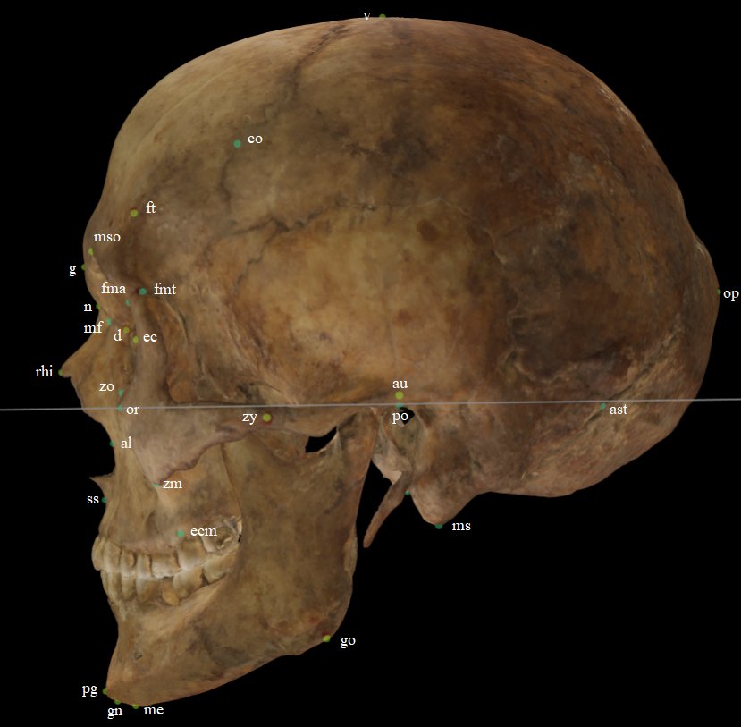 Fig. 1. Craniometric landmarks on a skull 3D model in frontal/lateral view.